The kinetochore is a protein complex assembled on the centromere region of a chromosome, essential for microtubule attachment during cell division. The centromere is the specialized DNA sequence that serves as the foundation for kinetochore formation and ensures proper chromosome segregation. Explore deeper into their distinct yet interdependent roles in mitosis and meiosis for a comprehensive understanding.
Main Difference
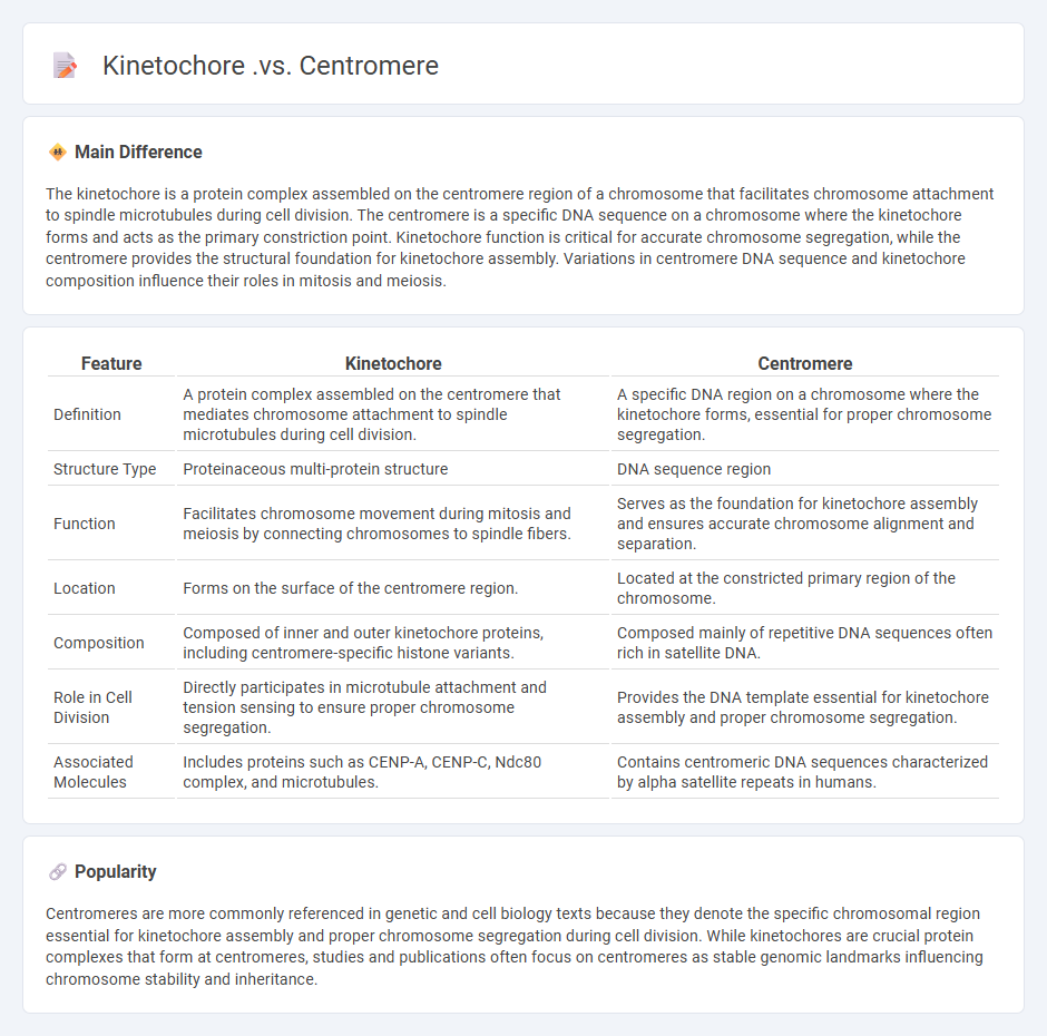
The kinetochore is a protein complex assembled on the centromere region of a chromosome that facilitates chromosome attachment to spindle microtubules during cell division. The centromere is a specific DNA sequence on a chromosome where the kinetochore forms and acts as the primary constriction point. Kinetochore function is critical for accurate chromosome segregation, while the centromere provides the structural foundation for kinetochore assembly. Variations in centromere DNA sequence and kinetochore composition influence their roles in mitosis and meiosis.
Connection
The kinetochore is a protein structure assembled on the centromere, serving as the attachment site for spindle microtubules during cell division. The centromere consists of specific DNA sequences and epigenetic markers that define the kinetochore assembly region. This connection ensures accurate chromosome segregation by facilitating the physical linkage between chromosomes and the mitotic spindle apparatus.
Comparison Table
| Feature | Kinetochore | Centromere |
|---|---|---|
| Definition | A protein complex assembled on the centromere that mediates chromosome attachment to spindle microtubules during cell division. | A specific DNA region on a chromosome where the kinetochore forms, essential for proper chromosome segregation. |
| Structure Type | Proteinaceous multi-protein structure | DNA sequence region |
| Function | Facilitates chromosome movement during mitosis and meiosis by connecting chromosomes to spindle fibers. | Serves as the foundation for kinetochore assembly and ensures accurate chromosome alignment and separation. |
| Location | Forms on the surface of the centromere region. | Located at the constricted primary region of the chromosome. |
| Composition | Composed of inner and outer kinetochore proteins, including centromere-specific histone variants. | Composed mainly of repetitive DNA sequences often rich in satellite DNA. |
| Role in Cell Division | Directly participates in microtubule attachment and tension sensing to ensure proper chromosome segregation. | Provides the DNA template essential for kinetochore assembly and proper chromosome segregation. |
| Associated Molecules | Includes proteins such as CENP-A, CENP-C, Ndc80 complex, and microtubules. | Contains centromeric DNA sequences characterized by alpha satellite repeats in humans. |
Chromosome Segregation
Chromosome segregation is a critical process during cell division where replicated chromosomes are evenly distributed into two daughter cells, ensuring genetic stability. This process primarily occurs during mitosis and meiosis, involving complex mechanisms like spindle fiber attachment to kinetochores and the action of motor proteins. Errors in chromosome segregation can lead to aneuploidy, which is associated with diseases such as Down syndrome and various cancers. Key proteins involved include cohesins, separase, and the spindle assembly checkpoint components that regulate accurate chromosome alignment and separation.
Centromere Structure
Centromeres are essential chromosome regions responsible for accurate chromosome segregation during cell division, characterized by repetitive DNA sequences and specialized nucleoprotein complexes. The core centromere contains alpha-satellite DNA in humans, which recruits kinetochore proteins such as CENP-A, a histone H3 variant critical for centromere identity and function. Centromere structure varies across species, with point centromeres in yeast and regional centromeres in most eukaryotes, reflecting differences in DNA sequence length and organization. Precise centromere architecture ensures proper spindle attachment and prevents chromosomal instability, a key factor in genetic disorders and cancer development.
Kinetochore Function
The kinetochore is a protein complex essential for chromosome segregation during cell division in eukaryotic cells. It assembles on the centromere of each chromosome, providing attachment sites for spindle microtubules that pull sister chromatids apart during mitosis and meiosis. Kinetochore function involves precise regulation of microtubule dynamics and tension sensing to ensure accurate chromosome alignment and segregation. Defects in kinetochore components can lead to aneuploidy, a hallmark of many cancers and genetic disorders.
Microtubule Attachment
Microtubule attachment is a critical process in cell division, ensuring proper chromosome alignment and segregation. Kinetochores, protein complexes assembled on centromeres, mediate the stable attachment of microtubules to chromosomes during mitosis. Errors in microtubule attachment can lead to aneuploidy, contributing to cancer development and other genetic disorders. Research on microtubule dynamics and kinetochore function provides insights into therapeutic targets for mitotic inhibitors in cancer treatment.
Mitotic Spindle
The mitotic spindle is a dynamic, microtubule-based structure essential for accurate chromosome segregation during mitosis in eukaryotic cells. Composed primarily of microtubules, centrosomes, and associated proteins, it organizes and separates sister chromatids to opposite poles of the cell. Key proteins such as kinesin and dynein motor proteins regulate spindle assembly and chromosome movement, ensuring proper cell division. Defects in mitotic spindle function can lead to aneuploidy and are implicated in cancer progression.
Source and External Links
Difference Between Centromere and Kinetochore - GeeksforGeeks - The centromere is the specific DNA region of the chromosome that holds two sister chromatids together, visible under a light microscope, while the kinetochore is a protein complex assembled on the centromere, visible only by electron microscopy, serving as the attachment site for spindle microtubules during cell division.
Plasticity in centromere organization and kinetochore composition - PMC - The centromere is the chromosomal region that acts as a platform for kinetochore assembly, and the kinetochore is the macromolecular complex that mediates the interaction between chromosomes and spindle microtubules to ensure proper chromosome segregation.
Functions of the centromere and kinetochore in chromosome ... - PMC - The centromere directs the assembly of the kinetochore, which forms specifically in mitosis to attach chromosomes to spindle microtubules, generate tension, and control checkpoint signaling for accurate chromosome segregation.
FAQs
What is a centromere?
A centromere is the essential chromosome region that connects sister chromatids and facilitates their proper segregation during cell division.
What is a kinetochore?
A kinetochore is a protein complex assembled on the centromere of a chromosome that attaches to spindle microtubules, facilitating chromosome segregation during cell division.
How are centromere and kinetochore different?
The centromere is a specific DNA sequence region on a chromosome where sister chromatids are most tightly connected, while the kinetochore is a protein complex assembled on the centromere that attaches chromosomes to spindle microtubules during cell division.
Where is the centromere located on a chromosome?
The centromere is located near the middle region of a chromosome, serving as the attachment site for spindle fibers during cell division.
What is the function of the kinetochore?
The kinetochore functions as the protein structure on chromatids where spindle fibers attach during cell division to ensure accurate chromosome segregation.
How do kinetochores interact with spindle fibers?
Kinetochores attach to spindle fibers by binding to microtubules, facilitating chromosome movement during cell division.
Why are both centromere and kinetochore important during cell division?
Centromeres ensure accurate chromosome segregation by serving as attachment sites for kinetochores, which mediate spindle fiber binding and chromosome movement during cell division.