Hemoptysis refers to the expectoration of blood originating from the lower respiratory tract, often associated with conditions like bronchitis, tuberculosis, or lung cancer. Hematemesis involves vomiting blood that primarily stems from gastrointestinal sources such as peptic ulcers, gastritis, or esophageal varices. Explore further to understand the distinguishing clinical features, diagnostic approaches, and treatment strategies for hemoptysis versus hematemesis.
Main Difference
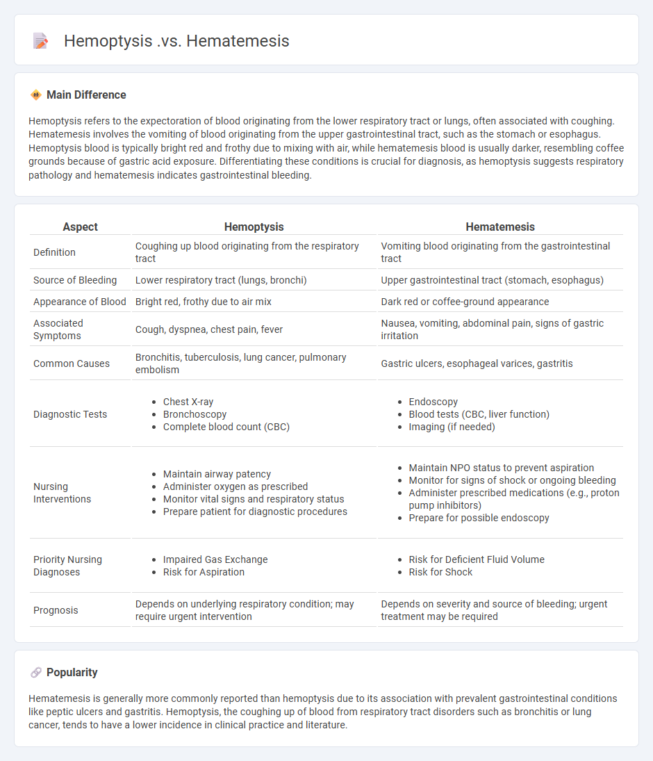
Hemoptysis refers to the expectoration of blood originating from the lower respiratory tract or lungs, often associated with coughing. Hematemesis involves the vomiting of blood originating from the upper gastrointestinal tract, such as the stomach or esophagus. Hemoptysis blood is typically bright red and frothy due to mixing with air, while hematemesis blood is usually darker, resembling coffee grounds because of gastric acid exposure. Differentiating these conditions is crucial for diagnosis, as hemoptysis suggests respiratory pathology and hematemesis indicates gastrointestinal bleeding.
Connection
Hemoptysis and hematemesis both involve the expulsion of blood but originate from different anatomical sources; hemoptysis is the coughing up of blood from the respiratory tract, while hematemesis is the vomiting of blood from the gastrointestinal tract. Conditions like severe coughing, lung infections, or pulmonary embolism can lead to hemoptysis, whereas gastroesophageal varices, peptic ulcers, or gastric erosions cause hematemesis. Differential diagnosis often requires imaging studies, endoscopy, and analysis of the blood's appearance to determine the precise source and underlying pathology.
Comparison Table
| Aspect | Hemoptysis | Hematemesis |
|---|---|---|
| Definition | Coughing up blood originating from the respiratory tract | Vomiting blood originating from the gastrointestinal tract |
| Source of Bleeding | Lower respiratory tract (lungs, bronchi) | Upper gastrointestinal tract (stomach, esophagus) |
| Appearance of Blood | Bright red, frothy due to air mix | Dark red or coffee-ground appearance |
| Associated Symptoms | Cough, dyspnea, chest pain, fever | Nausea, vomiting, abdominal pain, signs of gastric irritation |
| Common Causes | Bronchitis, tuberculosis, lung cancer, pulmonary embolism | Gastric ulcers, esophageal varices, gastritis |
| Diagnostic Tests |
|
|
| Nursing Interventions |
|
|
| Priority Nursing Diagnoses |
|
|
| Prognosis | Depends on underlying respiratory condition; may require urgent intervention | Depends on severity and source of bleeding; urgent treatment may be required |
Origin of Bleeding
Bleeding originates from damaged blood vessels when the vascular wall integrity is disrupted, causing blood to escape into surrounding tissues or external surfaces. It can be classified as arterial, venous, or capillary based on the vessel type involved, with arterial bleeding characterized by bright red, pulsatile flow due to high-pressure oxygenated blood. In nursing, assessing the severity of bleeding involves evaluating the source, volume, and rate, along with monitoring vital signs to prevent hypovolemic shock. Effective interventions include applying direct pressure, elevating the affected area, and using hemostatic agents or tourniquets when necessary to control hemorrhage.
Clinical Manifestations
Clinical manifestations in nursing refer to the observable signs and symptoms presented by patients that indicate underlying health conditions or diseases. These manifestations guide nurses in performing accurate assessments, developing effective care plans, and monitoring patient progress. Common examples include fever, pain, edema, and altered vital signs, which are critical for identifying acute or chronic pathologies. Understanding these clinical clues enhances early detection and improves patient outcomes through timely intervention.
Diagnostic Assessment
Diagnostic assessment in nursing involves systematically collecting and analyzing patient data to identify health problems and establish nursing diagnoses. It includes physical examinations, patient history, laboratory results, and diagnostic tests to determine the patient's condition accurately. Nurses use this information to develop individualized care plans that improve patient outcomes effectively. Accurate diagnostic assessments are critical for timely interventions in acute and chronic healthcare settings.
Nursing Interventions
Nursing interventions encompass a range of evidence-based actions designed to improve patient outcomes, including administering medications, monitoring vital signs, and providing emotional support. These interventions are guided by the nursing process, which involves assessment, diagnosis, planning, implementation, and evaluation. Effective nursing interventions reduce complications and promote recovery by addressing both physical and psychosocial needs of patients. Documentation of these interventions ensures continuity of care and supports interdisciplinary collaboration in healthcare settings.
Patient History Evaluation
Patient history evaluation in nursing is a systematic process of collecting comprehensive health information from patients to guide clinical decision-making. This assessment includes medical, surgical, family, and psychosocial histories, emphasizing chronic conditions such as diabetes, hypertension, and cardiac diseases. Accurate documentation and analysis of patient history enable early detection of health risks and personalized care planning. Skilled nurses use standardized tools like the HEADSS assessment for adolescents and the ROS (Review of Systems) to ensure thorough data collection.
Source and External Links
Hemoptysis Versus Hematemesis - Blog Details | Rigomo - Hemoptysis is coughing up bright red, frothy blood often with a tingling throat sensation, while hematemesis is vomiting dark red blood, often with nausea, upset stomach, and possible food particles; hematemesis blood is acidic and usually results in positive occult blood in stool, unlike hemoptysis blood which is neutral to alkaline.
Hemoptysis: Diagnosis and Management - AAFP - Hemoptysis is defined as spitting up blood from the lungs or bronchial tubes due to pulmonary bleeding, whereas hematemesis is vomiting of blood from the gastrointestinal tract; differentiating between them is critical for diagnosis and treatment.
Difference Between Hemoptysis and Hematemesis - Dr. Belal Bin Asaf - Hemoptysis involves coughing up blood originating from the respiratory tract, whereas hematemesis involves vomiting blood originating from the gastrointestinal tract.
FAQs
What is hemoptysis?
Hemoptysis is the expectoration of blood from the respiratory tract, indicating bleeding within the lungs or airways.
What is hematemesis?
Hematemesis is the vomiting of blood, indicating bleeding in the upper gastrointestinal tract.
What are the key differences between hemoptysis and hematemesis?
Hemoptysis is the coughing up of blood originating from the lower respiratory tract, often bright red and frothy, while hematemesis is the vomiting of blood from the upper gastrointestinal tract, typically darker and may contain food particles.
What causes hemoptysis?
Hemoptysis is caused by respiratory infections, bronchitis, pneumonia, tuberculosis, lung cancer, pulmonary embolism, bronchiectasis, trauma, or anticoagulant therapy.
What causes hematemesis?
Hematemesis is caused by upper gastrointestinal bleeding originating from conditions such as peptic ulcers, esophageal varices, gastritis, gastric cancer, or Mallory-Weiss tears.
How does the appearance of blood differ in hemoptysis and hematemesis?
Blood in hemoptysis is bright red and frothy due to oxygenation and air mixing, while blood in hematemesis is dark red or coffee-ground in appearance because of gastric acid exposure.
Which diagnostic tests are used for hemoptysis and hematemesis?
Chest X-ray, CT scan, bronchoscopy, and sputum analysis diagnose hemoptysis; upper endoscopy (esophagogastroduodenoscopy), abdominal ultrasound, and blood tests diagnose hematemesis.