Hypertonic solutions contain a higher concentration of solutes compared to the intracellular fluid, causing water to move out of cells and resulting in cell shrinkage. Hypotonic solutions have a lower solute concentration than the intracellular fluid, leading to water entering the cells and causing them to swell. Explore the detailed mechanisms and clinical applications of hypertonic and hypotonic solutions to enhance your understanding.
Main Difference
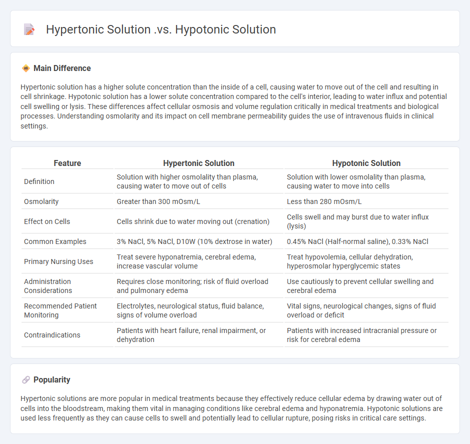
Hypertonic solution has a higher solute concentration than the inside of a cell, causing water to move out of the cell and resulting in cell shrinkage. Hypotonic solution has a lower solute concentration compared to the cell's interior, leading to water influx and potential cell swelling or lysis. These differences affect cellular osmosis and volume regulation critically in medical treatments and biological processes. Understanding osmolarity and its impact on cell membrane permeability guides the use of intravenous fluids in clinical settings.
Connection
Hypertonic and hypotonic solutions are connected by their relative solute concentrations compared to a cell's cytoplasm. A hypertonic solution has a higher solute concentration, causing water to move out of the cell, leading to cell shrinkage. In contrast, a hypotonic solution has a lower solute concentration, resulting in water moving into the cell and causing it to swell or burst.
Comparison Table
| Feature | Hypertonic Solution | Hypotonic Solution |
|---|---|---|
| Definition | Solution with higher osmolality than plasma, causing water to move out of cells | Solution with lower osmolality than plasma, causing water to move into cells |
| Osmolarity | Greater than 300 mOsm/L | Less than 280 mOsm/L |
| Effect on Cells | Cells shrink due to water moving out (crenation) | Cells swell and may burst due to water influx (lysis) |
| Common Examples | 3% NaCl, 5% NaCl, D10W (10% dextrose in water) | 0.45% NaCl (Half-normal saline), 0.33% NaCl |
| Primary Nursing Uses | Treat severe hyponatremia, cerebral edema, increase vascular volume | Treat hypovolemia, cellular dehydration, hyperosmolar hyperglycemic states |
| Administration Considerations | Requires close monitoring; risk of fluid overload and pulmonary edema | Use cautiously to prevent cellular swelling and cerebral edema |
| Recommended Patient Monitoring | Electrolytes, neurological status, fluid balance, signs of volume overload | Vital signs, neurological changes, signs of fluid overload or deficit |
| Contraindications | Patients with heart failure, renal impairment, or dehydration | Patients with increased intracranial pressure or risk for cerebral edema |
Osmolarity
Osmolarity measures the concentration of solutes in a fluid and is critical in nursing for maintaining proper fluid balance and preventing cellular dehydration or swelling. Accurate assessment of serum osmolarity, normally between 275-295 mOsm/kg, guides intravenous fluid therapy and electrolyte management in patients. Deviations from normal osmolarity levels can indicate conditions such as dehydration, hyponatremia, or hypernatremia, which require prompt intervention. Monitoring osmolarity supports optimal patient outcomes by ensuring homeostasis during critical care and routine nursing assessments.
Cell Volume Changes
Cell volume changes play a critical role in maintaining cellular homeostasis and are influenced by factors such as osmotic pressure, ion channel activity, and fluid balance. In nursing practice, understanding how conditions like dehydration, edema, and electrolyte imbalances affect cell volume helps in assessing patient status and guiding treatment. Techniques such as monitoring serum osmolarity and electrolyte levels provide valuable data for detecting abnormal cell swelling or shrinkage. Effective management of cell volume disturbances is essential to prevent complications like cellular dysfunction and organ failure.
Intravenous Fluid Types
Intravenous fluid types commonly used in nursing include crystalloids and colloids, each serving distinct clinical purposes. Crystalloids, such as normal saline (0.9% sodium chloride) and lactated Ringer's solution, are ideal for volume expansion and electrolyte replacement. Colloids like albumin and dextran contain larger molecules that remain in the vascular compartment longer, supporting oncotic pressure and hydration in cases of severe hypovolemia or hypoalbuminemia. Selection depends on patient condition, fluid balance, and electrolyte needs, guided by protocols to optimize outcomes in fluid therapy.
Indications and Contraindications
Nursing interventions are guided by clear indications that specify when certain treatments or procedures should be applied to improve patient outcomes, such as administering medications for infection control or providing wound care to promote healing. Contraindications highlight conditions or factors that render a treatment unsafe, including allergies to medications, unstable vital signs, or specific comorbidities like uncontrolled hypertension or bleeding disorders. Understanding these indications and contraindications ensures safe, effective nursing care tailored to individual patient needs documented in clinical guidelines like those from the American Nurses Association. Comprehensive assessment and ongoing monitoring are critical to adjusting nursing actions when patient status changes, preventing adverse events and optimizing recovery.
Clinical Effects and Risks
Clinical effects of nursing interventions significantly impact patient recovery, including improved wound healing, enhanced pain management, and reduced hospital stay duration. Nurses play a critical role in monitoring vital signs, administering medications, and preventing hospital-acquired infections, thereby decreasing morbidity rates. Risks in nursing practices include medication errors, needle-stick injuries, and exposure to infectious diseases, which can compromise both patient and nurse safety. Adherence to evidence-based protocols and continuous education minimizes these risks and optimizes patient outcomes.
Source and External Links
Difference Between Hypotonic and Hypertonic Solution - A hypotonic solution has less solute (and more solvent) than a cell, causing water to enter the cell and leading to cell swelling, while a hypertonic solution has more solute (and less solvent) than a cell, causing water to leave the cell and leading to cell shrinkage.
What is difference between hypotonic and hypertonic solution - In a hypotonic solution (external solute concentration lower than inside the cell), the cell swells as water flows in; in a hypertonic solution (external solute concentration higher than inside the cell), the cell shrinks as water flows out.
Tonicity: hypertonic, isotonic & hypotonic solutions (article) - A hypotonic solution causes water to move into the cell, increasing its volume, whereas a hypertonic solution causes water to move out of the cell, decreasing its volume.