Ecchymosis and petechiae are both types of bleeding under the skin but differ in size and appearance; ecchymosis typically appears as larger, bruise-like patches over 1 centimeter, while petechiae are tiny, pinpoint red or purple spots less than 3 millimeters. These skin manifestations result from capillary bleeding and can signify underlying medical conditions such as clotting disorders or vasculitis. Explore more to understand their causes, diagnosis, and treatment options.
Main Difference
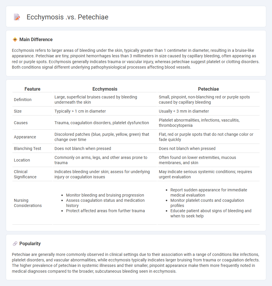
Ecchymosis refers to larger areas of bleeding under the skin, typically greater than 1 centimeter in diameter, resulting in a bruise-like appearance. Petechiae are tiny, pinpoint hemorrhages less than 3 millimeters in size caused by capillary bleeding, often appearing as red or purple spots. Ecchymosis generally indicates trauma or vascular injury, whereas petechiae suggest platelet or clotting disorders. Both conditions signal different underlying pathophysiological processes affecting blood vessels.
Connection
Ecchymosis and petechiae are both types of bleeding disorders characterized by blood leakage under the skin, resulting in visible discolorations. Ecchymosis refers to larger, bruise-like patches caused by blood pooling from damaged blood vessels, while petechiae are small, pinpoint red or purple spots caused by minor capillary bleeding. Both conditions often indicate underlying issues such as platelet dysfunction, coagulation abnormalities, or vascular fragility.
Comparison Table
| Feature | Ecchymosis | Petechiae |
|---|---|---|
| Definition | Large, superficial bruises caused by bleeding underneath the skin | Small, pinpoint, non-blanching red or purple spots caused by capillary bleeding |
| Size | Typically > 1 cm in diameter | Usually < 3 mm in diameter |
| Causes | Trauma, coagulation disorders, platelet dysfunction | Platelet abnormalities, infections, vasculitis, thrombocytopenia |
| Appearance | Discolored patches (blue, purple, yellow, green) that change over time | Flat, red or purple spots that do not change color or fade quickly |
| Blanching Test | Does not blanch when pressed | Does not blanch when pressed |
| Location | Commonly on arms, legs, and other areas prone to trauma | Often found on lower extremities, mucous membranes, and skin |
| Clinical Significance | Indicates bleeding under skin; assess for underlying injury or coagulation issues | May indicate serious systemic conditions; requires urgent evaluation |
| Nursing Considerations |
|
|
Ecchymosis
Ecchymosis refers to the discoloration of the skin caused by bleeding underneath, often resulting from trauma or medical conditions that affect blood clotting. In nursing, accurate assessment of ecchymosis includes noting its size, color changes, and location to monitor healing or identify underlying issues. Documenting ecchymosis helps differentiate between accidental injury and potential abuse, especially in vulnerable populations. Proper care involves protecting the affected area, managing pain, and addressing any contributing systemic conditions such as coagulation disorders or medication side effects.
Petechiae
Petechiae are small, pinpoint, non-blanching red or purple spots caused by capillary hemorrhage, commonly observed in patients with thrombocytopenia or clotting disorders. Nurses play a critical role in early detection by performing thorough skin assessments, especially in patients with hematologic conditions or those receiving anticoagulant therapy. Accurate documentation of the size, distribution, and progression of petechiae assists healthcare providers in diagnosing underlying causes such as sepsis, platelet dysfunction, or vasculitis. Prompt reporting and monitoring can improve patient outcomes by facilitating timely interventions and preventing complications.
Vascular fragility
Vascular fragility refers to the increased susceptibility of blood vessels to rupture or damage, often resulting in easy bruising or bleeding. In nursing care, understanding factors such as aging, corticosteroid use, and connective tissue disorders is essential for preventing injury and managing patient conditions. Nurses must monitor skin integrity closely and implement protective interventions to minimize trauma. Proper patient education on avoiding excessive pressure and monitoring for signs of bleeding is crucial in managing vascular fragility effectively.
Coagulation disorders
Coagulation disorders involve abnormalities in the blood clotting process that can lead to excessive bleeding or thrombosis. Common conditions include hemophilia, von Willebrand disease, and disseminated intravascular coagulation (DIC), each requiring specific nursing interventions to manage symptoms and prevent complications. Nurses play a critical role in monitoring coagulation profiles such as PT, aPTT, and platelet counts while administering anticoagulants or clotting factor replacements. Patient education on medication adherence, bleeding precautions, and recognizing signs of thrombosis is essential for improving outcomes in coagulation disorder management.
Skin assessment
Skin assessment in nursing involves a systematic examination of the skin's condition to identify signs of pressure ulcers, infections, dehydration, and other dermatological issues. Nurses use tools such as the Braden Scale to evaluate risk factors for skin breakdown and document skin color, moisture, temperature, and integrity. Accurate skin assessment supports early detection of complications, guiding timely interventions and improving patient outcomes. Consistent monitoring is crucial in vulnerable populations, including elderly patients and those with limited mobility.
Source and External Links
Discover the Difference Between Ecchymosis vs Petechiae - Ecchymosis are larger skin patches that appear blue, purple, or green and may be tender, while petechiae are tiny, flat red or purple spots resembling pinpricks, generally caused by capillary bleeding and unaffected by pressure.
Petechiation & Ecchymoses - Differential Diagnoses - Petechiae are small lesions under 3 mm from capillary bleeding, while ecchymoses are larger lesions caused by bleeding from small arteries or veins, both presenting as red to purple discolorations of skin or mucosa.
Petechia - Petechiae are pinpoint red or purple spots under 3 mm due to capillary hemorrhage, whereas ecchymoses are larger bruises exceeding 1 cm; these terms classify hemorrhages in different size categories.
FAQs
What are ecchymosis and petechiae?
Ecchymosis is a large bruise caused by bleeding under the skin, while petechiae are tiny, pinpoint red or purple spots resulting from minor bleeding from broken capillary blood vessels.
How do ecchymosis and petechiae differ in appearance?
Ecchymosis appears as large, irregularly shaped bruise-like patches, while petechiae are tiny, pinpoint red or purple spots usually less than 3 millimeters in diameter.
What causes ecchymosis?
Ecchymosis is caused by the leakage of blood from damaged blood vessels into the surrounding tissues, typically due to trauma, injury, or conditions affecting blood clotting.
What causes petechiae?
Petechiae are caused by capillary bleeding under the skin due to factors such as platelet deficiencies, blood clotting disorders, infections, physical trauma, or certain medications.
Are ecchymosis and petechiae dangerous?
Ecchymosis and petechiae can be dangerous if they indicate underlying conditions such as bleeding disorders, infections, or vascular diseases and require medical evaluation.
How are ecchymosis and petechiae diagnosed?
Ecchymosis and petechiae are diagnosed through clinical examination by identifying characteristic skin discolorations and confirmed with laboratory tests such as complete blood count, coagulation studies (PT, aPTT), platelet function tests, and sometimes skin biopsy to determine underlying causes.
When should you seek medical help for skin discoloration?
Seek medical help for skin discoloration if it appears suddenly, spreads rapidly, is accompanied by pain, swelling, itching, or fever, or if you notice persistent changes in color, shape, or size of a mole or lesion.